Microassay Thermal Denaturation of Ribonuclease A Probed by Circular Dichroism Spectroscopy
January 5, 2024
 Download This Application
Introduction
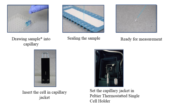
Circular dichroism (CD) is a useful technique to measure and analyze secondary structure and the thermal denaturation of proteins and nucleic acids in the far-UV region. Typically, a 1 mm pathlength rectangular cell is used for these measurements. However, this requires a sample volume of about 200 µL. In the case of samples where only very small volumes are available, JASCO now offers a new capillary cell for sample volumes smaller than 10 µL. The capillary cell also has a jacket for thermal ramping measurements, shown in Figure 1. CD measurements remain simple to perform and the capillary cells are inexpensive and disposable.
This application note demonstrates the use of a capillary cell and jacket for thermal ramping studies of ribonuclease A.
Experimental
| Measurement conditions | |
|---|---|
| Data acquisition interval | 0.2°C |
| Path length | 0.5 mm (capillary cell), 1 mm (rectangular cell) |
| Spectral bandwidth | 1 nm |
| Rising temperature rate | 1°C/min |
| Measurement wavelength | 222 nm |
| Response time | 8 sec |
1 mg/mL of ribonuclease A aqueous solution is drawn up into a capillary cell with a 0.5 mm optical pathlength, as seen in Figure 1. The cell is then inserted into the capillary jacket for thermal ramping CD measurements.
Keywords
210-CD-0025, Circular Dichroism, CD, capillary cell, microassay, thermal denaturation, ribonuclease A, Denatured Protein Analysis software, denaturation temperature, J-1500
Results
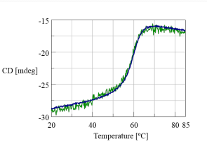
Figure 1 shows the thermal denaturation of ribonuclease A. The JASCO JWTDA-519 Denatured Protein Analysis software was used to calculate the denaturation temperature of ribonuclease A in both the capillary and rectangular cells. 59.4°C was calculated for the capillary cell which is in good agreement with 59.7°C obtained for the denaturation temperature in the rectangular cell.
Conclusion
This application note demonstrates that the denaturation temperature values, along with the thermal denaturation curves shown in Figure 2, illustrate that the microassay of ribonuclease A using the capillary cell and jacket can be carried out with high accuracy.
Note: The MSD-462 microsampling disc can be used for spectral scanning measurements on sample volumes between 2 and 10 µL. The MSD-462 applications are shown in the following Application Notes: 260-CD-0011 and 260-CD-0019.
Featured Products:
-
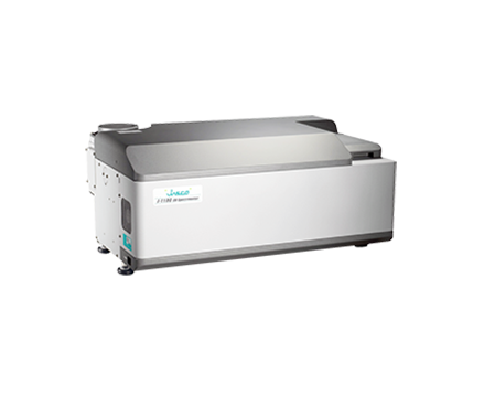
Automated high-throughput screening system for use with the J-1500/1700.
High-Throughput CD
-
The basic, compact model for research, QA/QC, and teaching applications.
J-1100
-
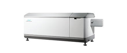
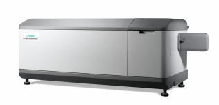
Highest performance with a wide range of accessories for maximum flexibility to meet complex research demands.
J-1500
-
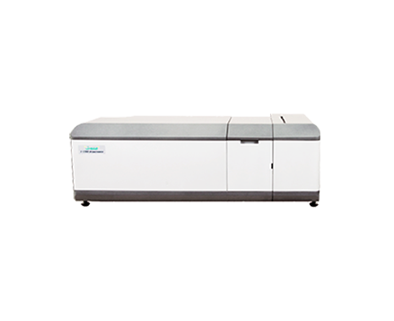
UV/Visible/NIR measurements up to 2,500 nm for MCD and specialized applications.
J-1700

Microassay Thermal Denaturation of Ribonuclease A Probed by Circular Dichroism Spectroscopy
Introduction
Circular dichroism (CD) is a useful technique to measure and analyze secondary structure and the thermal denaturation of proteins and nucleic acids in the far-UV region. Typically, a 1 mm pathlength rectangular cell is used for these measurements. However, this requires a sample volume of about 200 µL. In the case of samples where only very small volumes are available, JASCO now offers a new capillary cell for sample volumes smaller than 10 µL. The capillary cell also has a jacket for thermal ramping measurements, shown in Figure 1. CD measurements remain simple to perform and the capillary cells are inexpensive and disposable.
This application note demonstrates the use of a capillary cell and jacket for thermal ramping studies of ribonuclease A.
Experimental
| Measurement conditions | |
|---|---|
| Data acquisition interval | 0.2°C |
| Path length | 0.5 mm (capillary cell), 1 mm (rectangular cell) |
| Spectral bandwidth | 1 nm |
| Rising temperature rate | 1°C/min |
| Measurement wavelength | 222 nm |
| Response time | 8 sec |
1 mg/mL of ribonuclease A aqueous solution is drawn up into a capillary cell with a 0.5 mm optical pathlength, as seen in Figure 1. The cell is then inserted into the capillary jacket for thermal ramping CD measurements.
Results
Figure 1 shows the thermal denaturation of ribonuclease A. The JASCO JWTDA-519 Denatured Protein Analysis software was used to calculate the denaturation temperature of ribonuclease A in both the capillary and rectangular cells. 59.4°C was calculated for the capillary cell which is in good agreement with 59.7°C obtained for the denaturation temperature in the rectangular cell.
Conclusion
This application note demonstrates that the denaturation temperature values, along with the thermal denaturation curves shown in Figure 2, illustrate that the microassay of ribonuclease A using the capillary cell and jacket can be carried out with high accuracy.
Note: The MSD-462 microsampling disc can be used for spectral scanning measurements on sample volumes between 2 and 10 µL. The MSD-462 applications are shown in the following Application Notes: 260-CD-0011 and 260-CD-0019.
Keywords
210-CD-0025, Circular Dichroism, CD, capillary cell, microassay, thermal denaturation, ribonuclease A, Denatured Protein Analysis software, denaturation temperature, J-1500