Nanospectroscopy using a Near-Field Scanning Microspectrometer
August 25, 2022
 Download This Application
Introduction
Recent years have seen explosive growth in the use of organic materials, composites, or organics bound to an inorganic substrate, to solve engineering problems that traditionally were approached only with inorganics. The remarkable speed with which these new technologies are appearing is contrasted by the remarkable scarcity of non-destructive, invitro, and in-vivo techniques for characterizing the biophysical, chemical, and mechanical properties of these often delicate and nano-structured materials. Near-field scanning optical microscopy (NSOM) for quantitative evaluation of surfaces is a practical solution for this type of evaluation.
Traditional spectroscopy methods utilize ?far-field? light easily propagated through long distances. However, far field light is subject to diffraction effects, whether mirrors or lenses are used. Diffraction effects limit the amount of ?focus? that can be achieved, thus the spatial resolution for traditional microspectroscopy systems is limited to the wavelengths used; i.e. 10 microns for IR, ~1 micron for visible wavelengths.
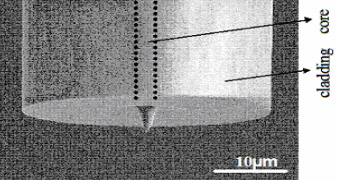
Near-field scanning optical microscopy (NSOM) is a type of microscopy where a light source with sub-wavelength spatial resolution is used as a scanning probe. The probe is scanned a few nanometers above the sample surface, the piezo-electric stage manipulators maintaining the probe above the surface using a feedback circuit from the probe electronics. A small aperture on the end of a tapered and gold-coated optical fiber is used as the near-field probe (Fig. 1). By illuminating a sample with the “near-field” created by the probe aperture, optical images can be constructed with a spatial resolution well beyond the usual “diffraction limit”, typically about 50 nm or greater.
Results
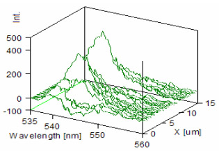
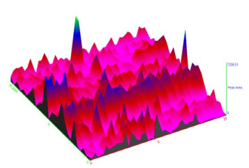
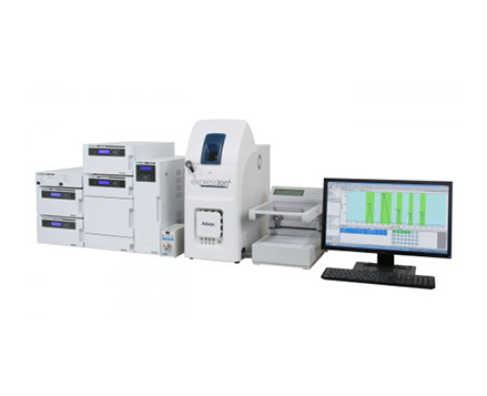
Luminescence spectra of nanotubes on a silica surface were collected using an NFS-310 near-field spectrometer. A near-field probe with an aperture of 50 nm was used to illuminate the sample with a 532 nm laser coupled to an optical fiber. An area of 15 x 20 microns was mapped to produce the spectra in Figure 2. Integration of the photoluminescence spectra was used to produce the intensity map in Figure 3. The luminescence intensities associated with discrete nanotubes corresponding to the ?peaks? displayed in the intensity map (Fig. 3).
Conclusion
Near-field scanning optical microscopy offers the ability to produce spectral data with subwavelength spatial resolution, beyond the diffraction limits of traditional spectroscopic microscopy methods.
Featured Products:
-

Compact fixed range mid-IR spectrometer with options to extend to the NIR/FIR
FT/IR-4X
-

SFC-4000 Analytical SFC for routine separation to method development.
Analytical SFC System
-
Single Quadrupole LC-MS

Nanospectroscopy using a Near-Field Scanning Microspectrometer
Introduction
Recent years have seen explosive growth in the use of organic materials, composites, or organics bound to an inorganic substrate, to solve engineering problems that traditionally were approached only with inorganics. The remarkable speed with which these new technologies are appearing is contrasted by the remarkable scarcity of non-destructive, invitro, and in-vivo techniques for characterizing the biophysical, chemical, and mechanical properties of these often delicate and nano-structured materials. Near-field scanning optical microscopy (NSOM) for quantitative evaluation of surfaces is a practical solution for this type of evaluation.
Traditional spectroscopy methods utilize ?far-field? light easily propagated through long distances. However, far field light is subject to diffraction effects, whether mirrors or lenses are used. Diffraction effects limit the amount of ?focus? that can be achieved, thus the spatial resolution for traditional microspectroscopy systems is limited to the wavelengths used; i.e. 10 microns for IR, ~1 micron for visible wavelengths.
Near-field scanning optical microscopy (NSOM) is a type of microscopy where a light source with sub-wavelength spatial resolution is used as a scanning probe. The probe is scanned a few nanometers above the sample surface, the piezo-electric stage manipulators maintaining the probe above the surface using a feedback circuit from the probe electronics. A small aperture on the end of a tapered and gold-coated optical fiber is used as the near-field probe (Fig. 1). By illuminating a sample with the “near-field” created by the probe aperture, optical images can be constructed with a spatial resolution well beyond the usual “diffraction limit”, typically about 50 nm or greater.
Results
Luminescence spectra of nanotubes on a silica surface were collected using an NFS-310 near-field spectrometer. A near-field probe with an aperture of 50 nm was used to illuminate the sample with a 532 nm laser coupled to an optical fiber. An area of 15 x 20 microns was mapped to produce the spectra in Figure 2. Integration of the photoluminescence spectra was used to produce the intensity map in Figure 3. The luminescence intensities associated with discrete nanotubes corresponding to the ?peaks? displayed in the intensity map (Fig. 3).
Conclusion
Near-field scanning optical microscopy offers the ability to produce spectral data with subwavelength spatial resolution, beyond the diffraction limits of traditional spectroscopic microscopy methods.