Detection of Amino Acids Using HPLC with CD Detection
November 17, 2022
Introduction
Amino acids are the “building blocks” of the body. Besides building cells and repairing tissue, they form antibodies to combat bacteria and viruses; they are part of the enzyme & hormonal system; they build nucleoproteins (RNA & DNA); they carry oxygen throughout the body and participate in muscle activity. The twenty naturally occurring amino acids that comprise proteins are almost all of the L- form. The L (Levorotatory) form is the stereoisomer that rotates plane polarized light to the left. One method to detect amino acids and determine their stereoisomers is with HPLC.
Click here to find out more about the CD detector
A tetrahedral carbon atom with 4 distinct constituents is said to be chiral. Chirality describes the ability of a molecule to rotate the plane of polarized light either to the right (dextrorotatory) or to the left (levorotatory). All of the amino acids in proteins exhibit the same absolute steric configuration. Therefore, they are all L-a-amino acids. D-amino acids are not found in proteins, although they exist in nature. The D-amino acids are found in polypetide antibiotics.
Table 1. The Amino Acids
| Amino Acid | Symbol | pK1 (COOH) | pK2 (NH2) |
|---|---|---|---|
| Glycine | Gly - G | 2.4 | 9.8 |
| Alanine | Ala - A | 2.4 | 9.9 |
| Valine | Val - V | 2.2 | 9.7 |
| Leucine | Leu - L | 2.3 | 9.7 |
| Serine | Ser-S | 2.2 | 9.2 |
| Threonine | Thr - T | 2.1 | 9.1 |
| Cysteine | Cys - C | 1.9 | 10.8 |
| Methionine | Met - M | 2.1 | 9.3 |
| Aspartic Acid | Asp - D | 2.0 | 9.9 |
| Asparagine | Asn - N | 2.1 | 8.8 |
| Asparagine | Asn - N | 2.1 | 8.8 |
| Glutamic Acid | Glu - E | 2.1 | 9.5 |
| Glutamine | Gln - Q | 2.2 | 9.1 |
| Arginine | Arg - R | 1.8 | 9.0 |
| Lysine | Lys - K | 2.2 | 9.2 |
| Histidine | His - H | 1.8 | 9.2 |
| Phenylalanine | Phe - F | 2.2 | 9.2 |
| Tyrosine | Tyr - Y | 2.2 | 9.1 |
| Tryptophan | Trp-W | 2.4 | 9.4 |
| Proline | Pro - P | 2.0 | 10.6 |
Peptides and polypeptides are polymers of alpha-amino acids. There are 20 α-amino acids that are relevant to the make-up of mammalian proteins. Several other amino acids are found in the body free or in combined states (i.e. not associated with peptides or proteins). These nonprotein associated amino acids perform specialized functions. Several of the amino acids found in proteins also serve functions distinct from the formation of peptides and proteins, e.g., tyrosine in the formation of thyroid hormones or glutamate as a neurotransmitter.
The role that amino acids play is crucial to the activity of many reactions. In solution, it is the nature of the amino acid R-groups that dictate structure-function relationships of peptides and proteins. The hydrophilic amino acids are generally found on the exterior of proteins as well as in the active centers of enzymatically active proteins. It is the amino acid R-groups that allow enzyme reactions to occur. Equally important is the ability of histidines in hemoglobin to buffer the H+ ions from carbonic acid ionization in red blood cells. It is this property of hemoglobin that allows it to exchange O2 and CO2 at the tissues or lungs, respectively. The aromatic R-groups in amino acids absorb ultraviolet light with an absorbance maximum in the range of 280nm. The ability of proteins to absorb ultraviolet light is predominantly due to the presence of the tryptophan which strongly absorbs ultraviolet light. This note will describe chiral detection of amino acids with HPLC.
Experimental
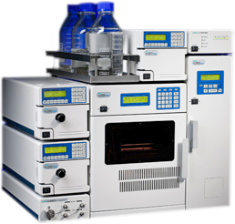
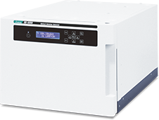
Several amino acids were examined using a Jasco HPLC system with a CD-2095 detector. The separation was accomplished with a CROWNPAK CR(+) (4.0 mm I.D. x 150 mm/L) column. The eluent was HClO4 with flow rates of 0.4 – 0.8 mL/min. The column temperature varied from 0 – 25 °C with an injection volume of 10 µL (2 mg/mL, 20µg). Amino acids were also separated following precolumn derivatization with DABS-Cl. For these a CrestPak C18S column was used with a mixture of: A) 8mM Sodium Dihydrogenphosphate dihydrate in H2O with 4% DMF, and B): ACN. Temperature was 40°C, injection volume 10µl and a flow rate 1mL/min. The gradient is show below.
| Time (min) | A (%) | B (%) |
|---|---|---|
| 0 | 85 | 15 |
| 9 | 70 | 30 |
| 14 | 59 | 41 |
| 16 | 49 | 51 |
| 18 | 46 | 54 |
| 23 | 10 | 90 |
Results

Figures 1 and 2 show the HPLC separation of proline, serine, threonine, cysteine, lysine and arginine with precolumn derivatization. Each of the compounds evaluated had both a positive and negative peak in the CD verifying the prescence of both enantiomers. In the case of molecules like Isoleucine (not show) which have 2 chiral centers, two separate sets of peaks are found, one set for each chiral center.

The separation of cysteine, proline, histidine, leucine, threonine, and phenanthroline was accomplished following precolumn derivatization with DABS-Cl. In these cases pre-derivitization is required for detection of the isomers because a chiral column was not used.

Figures 3 and 4 show the separation of cysteine and proline. Using derivatization allows a more complete separation and sharpens the peaks. Regardless of whether you separate your peaks with a chiral column or with precolumn derivatization, Jasco’s CD detector can offer sensitive and selective detection of chiral compounds such as amino acids.

Featured Products:

Detection of Amino Acids Using HPLC with CD Detection
Introduction

Amino acids are the “building blocks” of the body. Besides building cells and repairing tissue, they form antibodies to combat bacteria and viruses; they are part of the enzyme & hormonal system; they build nucleoproteins (RNA & DNA); they carry oxygen throughout the body and participate in muscle activity. The twenty naturally occurring amino acids that comprise proteins are almost all of the L- form. The L (Levorotatory) form is the stereoisomer that rotates plane polarized light to the left. One method to detect amino acids and determine their stereoisomers is with HPLC.
Click here to find out more about the CD detector
A tetrahedral carbon atom with 4 distinct constituents is said to be chiral. Chirality describes the ability of a molecule to rotate the plane of polarized light either to the right (dextrorotatory) or to the left (levorotatory). All of the amino acids in proteins exhibit the same absolute steric configuration. Therefore, they are all L-a-amino acids. D-amino acids are not found in proteins, although they exist in nature. The D-amino acids are found in polypetide antibiotics.
Table 1. The Amino Acids
| Amino Acid | Symbol | pK1 (COOH) | pK2 (NH2) |
|---|---|---|---|
| Glycine | Gly - G | 2.4 | 9.8 |
| Alanine | Ala - A | 2.4 | 9.9 |
| Valine | Val - V | 2.2 | 9.7 |
| Leucine | Leu - L | 2.3 | 9.7 |
| Serine | Ser-S | 2.2 | 9.2 |
| Threonine | Thr - T | 2.1 | 9.1 |
| Cysteine | Cys - C | 1.9 | 10.8 |
| Methionine | Met - M | 2.1 | 9.3 |
| Aspartic Acid | Asp - D | 2.0 | 9.9 |
| Asparagine | Asn - N | 2.1 | 8.8 |
| Asparagine | Asn - N | 2.1 | 8.8 |
| Glutamic Acid | Glu - E | 2.1 | 9.5 |
| Glutamine | Gln - Q | 2.2 | 9.1 |
| Arginine | Arg - R | 1.8 | 9.0 |
| Lysine | Lys - K | 2.2 | 9.2 |
| Histidine | His - H | 1.8 | 9.2 |
| Phenylalanine | Phe - F | 2.2 | 9.2 |
| Tyrosine | Tyr - Y | 2.2 | 9.1 |
| Tryptophan | Trp-W | 2.4 | 9.4 |
| Proline | Pro - P | 2.0 | 10.6 |
Peptides and polypeptides are polymers of alpha-amino acids. There are 20 α-amino acids that are relevant to the make-up of mammalian proteins. Several other amino acids are found in the body free or in combined states (i.e. not associated with peptides or proteins). These nonprotein associated amino acids perform specialized functions. Several of the amino acids found in proteins also serve functions distinct from the formation of peptides and proteins, e.g., tyrosine in the formation of thyroid hormones or glutamate as a neurotransmitter.
The role that amino acids play is crucial to the activity of many reactions. In solution, it is the nature of the amino acid R-groups that dictate structure-function relationships of peptides and proteins. The hydrophilic amino acids are generally found on the exterior of proteins as well as in the active centers of enzymatically active proteins. It is the amino acid R-groups that allow enzyme reactions to occur. Equally important is the ability of histidines in hemoglobin to buffer the H+ ions from carbonic acid ionization in red blood cells. It is this property of hemoglobin that allows it to exchange O2 and CO2 at the tissues or lungs, respectively. The aromatic R-groups in amino acids absorb ultraviolet light with an absorbance maximum in the range of 280nm. The ability of proteins to absorb ultraviolet light is predominantly due to the presence of the tryptophan which strongly absorbs ultraviolet light. This note will describe chiral detection of amino acids with HPLC.
Experimental

Several amino acids were examined using a Jasco HPLC system with a CD-2095 detector. The separation was accomplished with a CROWNPAK CR(+) (4.0 mm I.D. x 150 mm/L) column. The eluent was HClO4 with flow rates of 0.4 – 0.8 mL/min. The column temperature varied from 0 – 25 °C with an injection volume of 10 µL (2 mg/mL, 20µg). Amino acids were also separated following precolumn derivatization with DABS-Cl. For these a CrestPak C18S column was used with a mixture of: A) 8mM Sodium Dihydrogenphosphate dihydrate in H2O with 4% DMF, and B): ACN. Temperature was 40°C, injection volume 10µl and a flow rate 1mL/min. The gradient is show below.
| Time (min) | A (%) | B (%) |
|---|---|---|
| 0 | 85 | 15 |
| 9 | 70 | 30 |
| 14 | 59 | 41 |
| 16 | 49 | 51 |
| 18 | 46 | 54 |
| 23 | 10 | 90 |
Results

Figures 1 and 2 show the HPLC separation of proline, serine, threonine, cysteine, lysine and arginine with precolumn derivatization. Each of the compounds evaluated had both a positive and negative peak in the CD verifying the prescence of both enantiomers. In the case of molecules like Isoleucine (not show) which have 2 chiral centers, two separate sets of peaks are found, one set for each chiral center.

The separation of cysteine, proline, histidine, leucine, threonine, and phenanthroline was accomplished following precolumn derivatization with DABS-Cl. In these cases pre-derivitization is required for detection of the isomers because a chiral column was not used.

Figures 3 and 4 show the separation of cysteine and proline. Using derivatization allows a more complete separation and sharpens the peaks. Regardless of whether you separate your peaks with a chiral column or with precolumn derivatization, Jasco’s CD detector can offer sensitive and selective detection of chiral compounds such as amino acids.


 Download This Application