Enhanced Observation in FTIR Microscopy
April 25, 2024
 Download This Application
Introduction
IR microscopes use several different options for sample observation. In the simplest mode a measurement cassegrain is also used for sample viewing, in most cases this is sufficient for identifying the measurement location. However, in some instance the sample may lack detail, and more enhanced observation modes may be required. The IRT-5000 and IRT-7000 both benefit from a multi-position objective turret on which a choice of cassegrains may be mounted, ATRs with view-through observation for direct sample viewing and a refractive objective to act more like a light microscope. The refractive objective can be used to locate and identify a location with more detail prior to measurement using the cassegrain objective or ATR.
The IRT-5000 and IRT-7000 include a high resolution CMOS camera with 3x optical zoom (and a digital zoom) for the highest clarity sample observation. For enhanced observation, several optional measurement modes are available, each one can provide improved viewing of samples depending on the properties of the sample being analyzed.
Fluorescence Observation
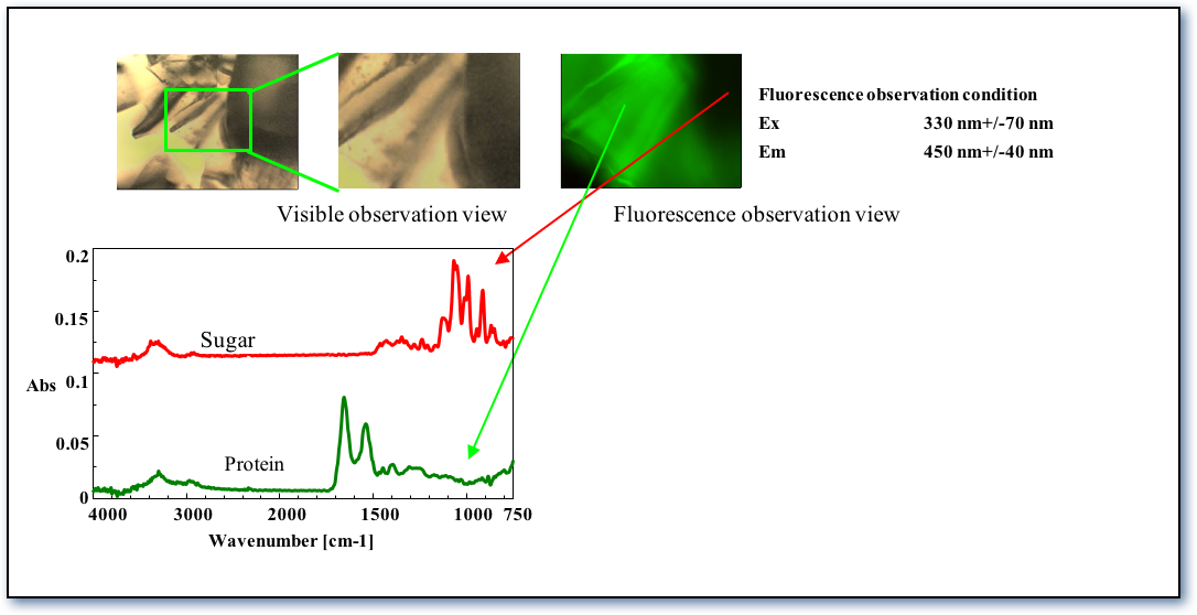
Fluorescence is a useful technique for differentiating a sample location for measurement, especially where they look similar using simple white light illumination. By exploiting the fluorescent properties it may be easier to see the differences prior to measurement. In the example below a powder sample that could not be easily identified using visible observation could be seen using the fluorescence observation accessory (as shown in Figure 1) a region in a mixed protein/sugar sample could be observed fluorescing in green. Infrared spectra were measured in both the green and black areas. Protein was identified in the green area, and sugar was found in the black area.
Polarization Observation
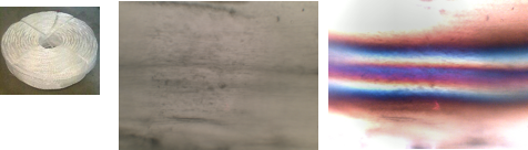
The use of polarized light in optical microscopy is well documented, birefringent materials can benefit significantly from polarized light to enhance contrast. In the example below a stretched vinyl sample was used (Figure 2), the stretched area which has very little contrast under simple un-polarized white light can be seen in a number of strata when viewed with polarized light. Polarization is extremely useful for observing samples with orientated structures.
Differential Interference Observation
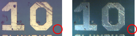
Differential Interference Contrast (DIC) uses two orthogonally polarized light beams, which interfere to provide contrast in objects that appear flattened when observed with simple unpolarized white light. It can be applied to biological and non-biological samples. In the example below, a printed circuit board was measured using standard observation and DIC observation; uneven points and scratches on the sample were more clearly observed using DIC.
Keywords
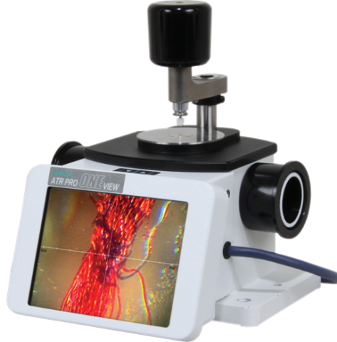
280-SO-0008, ATR PRO ONE, Spectrometers
Featured Products:
-
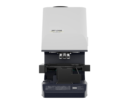
The IRT-5000 is an innovative FTIR microscope with powerful sample identification and imaging.
IRT-5000 Series FTIR Microscopes
-
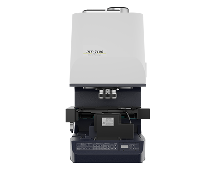
The IRT-7000 16-element linear array microscope for fast imaging and kinetics.
IRT-7000 FTIR Microscope

Enhanced Observation in FTIR Microscopy
Introduction
IR microscopes use several different options for sample observation. In the simplest mode a measurement cassegrain is also used for sample viewing, in most cases this is sufficient for identifying the measurement location. However, in some instance the sample may lack detail, and more enhanced observation modes may be required. The IRT-5000 and IRT-7000 both benefit from a multi-position objective turret on which a choice of cassegrains may be mounted, ATRs with view-through observation for direct sample viewing and a refractive objective to act more like a light microscope. The refractive objective can be used to locate and identify a location with more detail prior to measurement using the cassegrain objective or ATR.
The IRT-5000 and IRT-7000 include a high resolution CMOS camera with 3x optical zoom (and a digital zoom) for the highest clarity sample observation. For enhanced observation, several optional measurement modes are available, each one can provide improved viewing of samples depending on the properties of the sample being analyzed.
Fluorescence Observation
Fluorescence is a useful technique for differentiating a sample location for measurement, especially where they look similar using simple white light illumination. By exploiting the fluorescent properties it may be easier to see the differences prior to measurement. In the example below a powder sample that could not be easily identified using visible observation could be seen using the fluorescence observation accessory (as shown in Figure 1) a region in a mixed protein/sugar sample could be observed fluorescing in green. Infrared spectra were measured in both the green and black areas. Protein was identified in the green area, and sugar was found in the black area.
Polarization Observation
The use of polarized light in optical microscopy is well documented, birefringent materials can benefit significantly from polarized light to enhance contrast. In the example below a stretched vinyl sample was used (Figure 2), the stretched area which has very little contrast under simple un-polarized white light can be seen in a number of strata when viewed with polarized light. Polarization is extremely useful for observing samples with orientated structures.
Differential Interference Observation
Differential Interference Contrast (DIC) uses two orthogonally polarized light beams, which interfere to provide contrast in objects that appear flattened when observed with simple unpolarized white light. It can be applied to biological and non-biological samples. In the example below, a printed circuit board was measured using standard observation and DIC observation; uneven points and scratches on the sample were more clearly observed using DIC.
Keywords
280-SO-0008, ATR PRO ONE, Spectrometers