Raman Imaging of Samples with Uneven or Rough Surfaces using Confocal Microscopy with Surface Scan Imaging (SSI)
April 25, 2024
 Download This Application
Introduction
 A confocal Raman microspectrometer is an excellent choice for analysis of materials with high spatial resolution – in the order of submicrons. This makes it possible to characterize and image small components such as APIs or bulking agents and foreign materials down to 1 μm, as well as extremely thin layers. However, to achieve high spatial resolution it is necessary to use objective lenses with high magnification. This results in a depth-of-field that is extremely narrow; any slight deviation in the focus at the measurement surface greatly affects to the intensity of the measured spectrum. Imaging measurement of a sample with an uneven or a sloping surface may result in poor measurement unless the objective lens is refocused at each measurement point and can lead to extremely long measurement times.
To make extremely fast measurement in a short time JASCO has developed Surface Scan Imaging (SSI). SSI is used to assess the surface shape of the sample as height information of the stage prior to measurement and Raman measurement is made while adjusting the stage Z axis according to this surface shape information. Using SSI, a sample with a very uneven surface can now be measured to give good Raman spectra with good S/N at all data points.
Keywords
260-AN-0017, Raman, Surface Scan Imaging, SSI, Confocal Raman Imaging
Results
Comparison of SSI with Conventional Autofocus Techniques
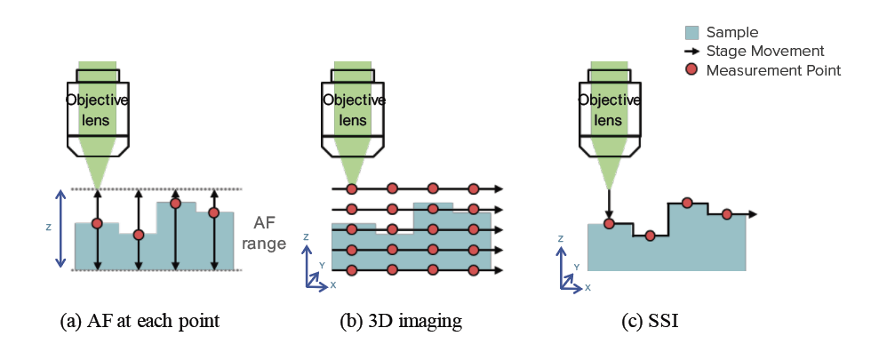
Conventionally, Raman imaging measurement of a sample with surface roughness was performed with autofocus (AF) was carried out at each measurement point (figure 1-a), or XYZ 3D imaging measurement was carried out (figure 1-b).
SSI (figure 1-c) calculates the sample height (irregularity) information from a multifocus* image and performs imaging measurement while scanning the stage based on this information. Although it also depends on measurement conditions and sample shape, the time required for Raman imaging measurement is greatly shortened compared with the conventional method.
* The multifocus image (all-focus image) is an image obtained when the stage is scanned in the Z direction to acquire multiple observation images with different focal points, and each layer that is in focus is extracted and a ‘single in-focus image’ is synthesized for the entire field of view. This multifocus image allow the details the entire surface shape of the sample to be identified prior to measurement.
Raman Imaging Measurement of a Pharmaceutical Tablet using SSI
Imaging analysis of the uneven part of the surface of a tablet (concave and convex difference: approximately 170 μm) was measured using SSI. The measurement parameters are as follows:
| Measurement Conditions | |
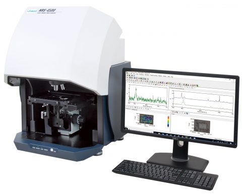
| Instrument | NRS-4500 Laser Raman Spectrometer |
| Excitation Wavelength | 532 nm |
| Objective Lens | 20x (long working distance type) |
| Interval | 10 µm |
| Measurement Points | 54 x 28 points |
| Exposure Time | 0.5 second |
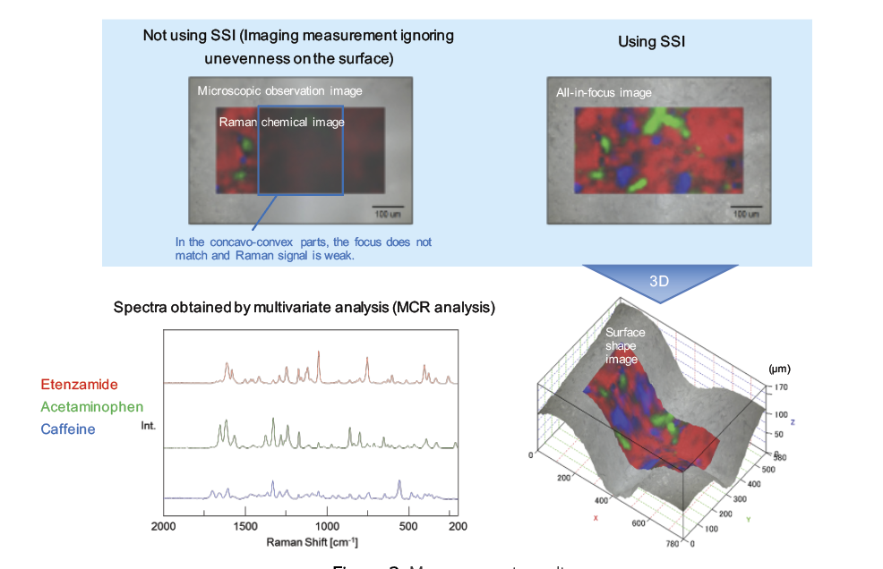
When measurement is performed following the surface shape by using SSI, a clear Raman chemical image can be obtained because the focus match at each point. By overlapping the Raman chemical image on the 3D surface shape image, the quantitative result and shape information of concave-convex part of the sample can be checked simultaneously.
Conclusion
Using SSI, Raman imaging measurement of a sample with surface unevenness and inclination can be performed easily and quickly. In addition to the tablet measurement shown here, SSI make it easier to analyze a wide range of samples with surface roughness including rock and concrete, DLC coating, resins, foods, medicines, fertilizer granules etc.
Featured Products:

Raman Imaging of Samples with Uneven or Rough Surfaces using Confocal Microscopy with Surface Scan Imaging (SSI)
Introduction
A confocal Raman microspectrometer is an excellent choice for analysis of materials with high spatial resolution – in the order of submicrons. This makes it possible to characterize and image small components such as APIs or bulking agents and foreign materials down to 1 μm, as well as extremely thin layers. However, to achieve high spatial resolution it is necessary to use objective lenses with high magnification. This results in a depth-of-field that is extremely narrow; any slight deviation in the focus at the measurement surface greatly affects to the intensity of the measured spectrum. Imaging measurement of a sample with an uneven or a sloping surface may result in poor measurement unless the objective lens is refocused at each measurement point and can lead to extremely long measurement times.
To make extremely fast measurement in a short time JASCO has developed Surface Scan Imaging (SSI). SSI is used to assess the surface shape of the sample as height information of the stage prior to measurement and Raman measurement is made while adjusting the stage Z axis according to this surface shape information. Using SSI, a sample with a very uneven surface can now be measured to give good Raman spectra with good S/N at all data points.
Results
Comparison of SSI with Conventional Autofocus Techniques
Conventionally, Raman imaging measurement of a sample with surface roughness was performed with autofocus (AF) was carried out at each measurement point (figure 1-a), or XYZ 3D imaging measurement was carried out (figure 1-b).
SSI (figure 1-c) calculates the sample height (irregularity) information from a multifocus* image and performs imaging measurement while scanning the stage based on this information. Although it also depends on measurement conditions and sample shape, the time required for Raman imaging measurement is greatly shortened compared with the conventional method.
* The multifocus image (all-focus image) is an image obtained when the stage is scanned in the Z direction to acquire multiple observation images with different focal points, and each layer that is in focus is extracted and a ‘single in-focus image’ is synthesized for the entire field of view. This multifocus image allow the details the entire surface shape of the sample to be identified prior to measurement.
Raman Imaging Measurement of a Pharmaceutical Tablet using SSI
Imaging analysis of the uneven part of the surface of a tablet (concave and convex difference: approximately 170 μm) was measured using SSI. The measurement parameters are as follows:
| Measurement Conditions | |
| Instrument | NRS-4500 Laser Raman Spectrometer |
| Excitation Wavelength | 532 nm |
| Objective Lens | 20x (long working distance type) |
| Interval | 10 µm |
| Measurement Points | 54 x 28 points |
| Exposure Time | 0.5 second |
When measurement is performed following the surface shape by using SSI, a clear Raman chemical image can be obtained because the focus match at each point. By overlapping the Raman chemical image on the 3D surface shape image, the quantitative result and shape information of concave-convex part of the sample can be checked simultaneously.
Conclusion
Using SSI, Raman imaging measurement of a sample with surface unevenness and inclination can be performed easily and quickly. In addition to the tablet measurement shown here, SSI make it easier to analyze a wide range of samples with surface roughness including rock and concrete, DLC coating, resins, foods, medicines, fertilizer granules etc.
Keywords
260-AN-0017, Raman, Surface Scan Imaging, SSI, Confocal Raman Imaging