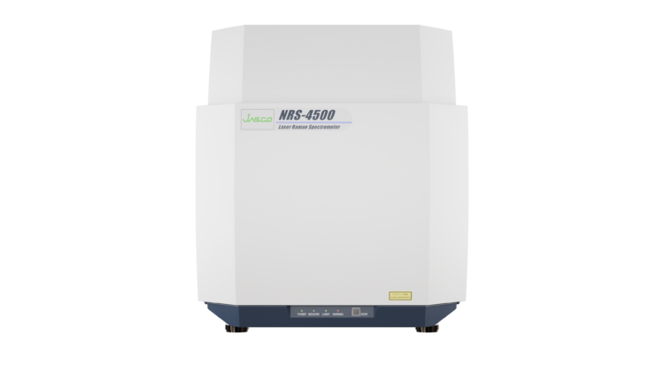


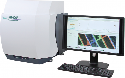
NRS-4500 Confocal Raman Microscope
The NRS-4500 Series is a purpose-designed Confocal Raman Microscope with a wide functionality to make setup fast and easy. When changing lasers the selection and alignment of the laser, rejection filter, and grating are fully automated. The laser spot is observed on all models including 1,064 nm ensuring that the sample being viewed is actually being measured. Sample identification, imaging analysis, and identification can all be done in real-time, speeding up the process of preparing the sample to seeing the results.
Optical Configuration
The f200 spectrograph includes up to 4 automatically selected gratings and detection by a high-performance CCD or InGaAs array (with options for electron multiplying EMCCDs). The observation system includes a super-resolution CMOS camera with Olympus optics and a choice of objectives from x5 to x100. Objective options include NIR and long working distance for working with heating cooling/sample stages.
Class 1 Laser Safety
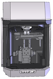
The standard Class 1 safety cabinet
Confocal Imaging
An automatic XYZ stage is used for confocal imaging with X/Y spatial resolution of 1 µm (Z=1.5 µm). Spectra Manager™ Suite with the Imaging Analysis application is used to collect a three-dimensional image of the sample which can be rotated and viewed from any angle. The image map is created using peak data corresponding to key functional groups and analysis of multiple peaks can be combined to create powerful data images.
Rigid Optical Bench and Laser Image
The Raman microscope has a completely rigid structure to prevent flexing and is not built around an optical microscope. Switching between observation and measurement modes is completely automatic and can be done with the safety cabinet closed.
Standard Configuration for a Raman Microscope
532 nm/785 nm Lasers with Matching Edge Filters
 The NRS-4500 Raman Microscope offers several standard configurations including the typical 633 nm single laser or 532 nm/785 nm laser combination with matching notch or edge filters, with an option for a third laser which can be mounted internally or externally. Observation of the laser spot ensures perfect alignment of the target sample position and indicates surface roughness. All laser wavelengths are selected in the software and once selected, the optical system (including the laser) is automatically aligned for optimal throughput and resolution. Four software-selectable gratings control the spectral range and resolution from 8000 to 100 cm-1 (8000 to 50 cm-1 as an option). The grating direct drive system includes a rotary encoder to ensure excellent wavelength reproducibility ±0.2cm-1.
Fluorescence Rejection in a Raman Microscope
457nm Laser and Patented Fluorescence Rejection Algorithm
JASCO has developed new and novel (patented) mechanisms to deal with sample fluorescence. As with other Raman systems, we can utilize laser wavelengths of 785 nm and up to 1,064 nm, but we have recently incorporated a 457 nm laser that offers a significantly higher Raman signal, improved spatial resolution, and much lower fluorescence for many different sample types. Selecting a different excitation laser wavelength is only one of the ways the NRS-4500 minimizes fluorescence interference. The Fluorescence Rejection algorithm (patented) included in the Spectra Manager™ can effectively remove or minimize fluorescence regardless of the laser wavelength used either at the time of measurement or during post-processing.
Flexible Sampling Options for a Raman Microscope
With a choice of refractive objectives, both micro and macro measurements are possible as well as options for long working distance objectives for heating/cooling stages and other sampling accessories. The NRS-4500 can be used with either a manual sample stage or the PC-controlled, automated XYZ confocal mapping stage. A fiber probe can be added for external measurement.
Powerful ‘UserAssist’ Control for Both New Users and Experienced Spectroscopists
The ‘UserAssist’ software aids the user in setting up the NRS-4500 for sample measurement; a simple sequence guide takes you through setup and optimization of measurement parameters with helpful advice and tips, such as a warning if you have the laser intensity set too high. When each of the parameters has been set, the NRS-4500 automatically selects the laser and matching notch filter, the grating for the appropriate resolution, focuses on the sample, and then the spectral measurement is performed.
‘Sample Search’ Function
The new ‘Sample Search’ function is used with the automated XYZ stage. A new algorithm developed by JASCO (patent pending) analyzes the microscopic image and automatically selects measurement position(s) based on the size, contrast, and/or color of the target material. The user simply clicks the measurement button to execute spectral measurements of the desired sample positions.
Chemical Image Identification and Functional Groups Registry
To provide faster Raman microscope image processing, the Imaging Analysis software includes a ‘Registry’ of possible functional groups or other relevant compound information based on peak height or area calculations. After peak height or area calculation is developed, it can be saved to the Registry for future analysis use. The registry includes the peak calculation information and a ‘label’ describing the relevant vibrational motion. Image maps can be developed from mapping data simply by clicking on the desired, registered calculation to obtain a false color image of the peak intensity data for the registered calculation.
6 Cool Features for the NRS-4500 Raman Microscope
- QRI (Quick Raman Imaging): High-speed imaging for large areas by combining a fast stage with an Electron Multiplying CCD (EMCCD) detector to take very quick exposures across the sample.
- Macro Measurement: A simple slide-on macro lens allows the user to measure liquids or powders in cuvettes or glass vials. Automatic spectral comparison indicates the compound in the sample.
- Analysis Wizard: Automatically go from viewing raw unprocessed data through all of the processing required to prepare every spectrum for multivariate curve regression to identify the principal components and image them using false color maps.
- SSI (Surface Scan Imaging) A fast and powerful tool for productivity when imaging or mapping a large sample area of a rough, uneven, or tilted surface, such as a geological sample or a pharmaceutical tablet.
- FocusNAV: The auto-focus function allows the Raman to do the focusing, not the user when surveying a sample for measurement.
- Advanced Sample Search: For the measurement of particulates, image analysis is applied to the visual image to identify and select particles based on size, shape, and contrast.