Fundamentals of Vibrational Spectroscopy
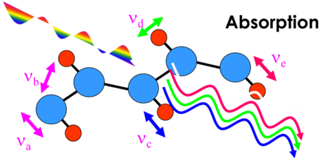
Vibrational spectroscopy is the study of the interaction between light and matter Incident white light interacts with all vibrational modes of the sample at once and emerges with characteristic missing lines. Monochromatic light passes through a molecule or crystal lattice and interacts with the naturally vibrating atomic bond system. Each bond has a characteristic vibrational frequency, n, and the light can emerge from the material either unchanged, or with a frequency that is shifted up or down by the bond vibration. Because we use monochromatic light, examination of the resulting spectrum directly yields the bond vibrational frequencies.
What are the fundamental differences and complementary aspects of Raman and IR absorption measurement?
Rayleigh scattered light undergoes no energy change. The cross-section for Raman scattering is very small- less than 10-6
Stokes scattering, the “normal” Raman peaks, loses energy: anti-Stokes scattering gains energy (anti-Stokes peaks usually much weaker)
Infrared is more sensitive to functional groups with very oriented dipole moments. But Raman is more sensitive to bonds that are equally shared. Generally, what is strong in the Raman is weak in the infrared and vice versa.
In simple, highly symmetric molecules such as benzene and CCl4, the Raman and infrared spectra can be mutually exclusive; some bands only present in the Raman and some only in the infrared. However, either spectrum fully describes the molecular vibrational activity.
Inorganics, with polar bonds, have few Raman active vibrations. In these materials, phonons (lattice vibrations) can dominate the Raman spectrum: we can study the solid-state structure with Raman spectroscopy. Some materials (silicon, diamond) characterized by a single sharp phonon mode.
Sample preparation for infrared spectroscopy is critical and often tedious. There is no sample preparation for Raman samples. Infrared samples require fragile, water-sensitive sample cells or complex, expensive sampling accessories with limited spectral ranges. Raman samples are held in a quartz cuvette or otherwise placed in the path of the laser. Water is a nuisance for most infrared analyses. Water is an ideal solvent for Raman sampling, with a very weak interference.
In spectroscopy we generally use the red-shifted Stokes side of the spectra as these peaks are the strongest, however the spectra are qualitatively symmetrical
Raman scattering directly probes the electronic and lattice dynamic properties of materials, giving us, both readily interpreted “fingerprint” spectra, and a wealth of more detailed physical information
When would you select one technique in preference to the other?
Can be used to analyze a wide variety of samples – liquids, powders, solids and solutions
Either technique will provide identification of unknowns, confirmation of molecular structure and quantitative analysis of mixtures
Each method has advantages and disadvantages
Peaks in the low wavenumber range are attributed to lattice vibrations (vibrations of atoms in crystals) and vibration modes between heavy atoms. For example, it is possible to check the difference in the crystal structure of calcium carbonate and the difference between quartz and glass.
As vibrational techniques they are quite insensitive, what can you do to increase sensitivity and improve dynamic range
Infrared quantitation can approach ppm levels for some samples and even ppb levels for gases, but most samples are limited to detection levels in the 0.01 to 0.001% range.
Raman spectra can be used for quantitation with a detection limit approaching 0.01% in some cases.